
X-ray imaging opens door to nanopaleomagnetism of Fe-Ni meteorites
We are pleased to announce the publication of our latest open access paper in Earth and Planetary Science Letters:
Bryson J, Herrero-Albillos J, Kronast F, Ghidini M, Redfern SAT, van der Laan G, and Harrison RJ (2014) Nanopaleomagnetism of meteoritic Fe–Ni studied using X-ray photoemission electron microscopy. Earth and Planetary Science Letters 396, 125–133.
This paper is the second in our series on the Tazewell IIICD Fe-Ni meteorite. Our first paper (open access download) described the fundamental chemical and magnetic structure of the cloudy zone, as revealed by electron holography, and put forward the idea that cloudy zones could contain a continuous, time-resolved record of the magnetic field generated by asteroidal dynamos. Our latest paper takes a giant step towards this goal by demonstrating that nanoscale paleomagnetic information can be extracted from the cloudy zone using X-ray Photoemission Electron Microscopy (XPEEM). Our results reveal in unprecedented detail the magnetic structure of the Widmanstätten sequence (kamacite, tetrataenite rim, cloudy zone, plessite). Furthermore, we demonstrate a methodology for performing quantitative paleomagnetic analysis of XPEEM images, which shows the way forward for future paleomagnetic studies of Fe-Ni within meteorites (e.g. pallasites) that may have experienced a dynamo field on their parent body.
XPEEM works by illuminating a polished surface with a beam of circularly polarised X-rays, tuned to the absorption edge of the element of interest (Fe L2,3 in this case). Photo electrons emitted from the surface are accelerated through a series of lenses to form an image of the sample with 30-40 nm resolution over a field-of-view of 5-20 µm. Magnetic contrast in the image is caused by XMCD (differential absorption of left versus right circularly polarised light), which produces a signal that is directly proportional to the magnetisation of the sample projected along the X-ray beam direction.
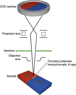
Schematic illustration of the XPEEM method.
The images below show the complex nanoscale magnetic structure associated with the cloudy zone – a spinodal intergrowth of magnetically hard (FeNi) and soft (Fe3Ni) phases. Regions of red and blue correspond to islands of FeNi magnetised along the three possible <100> easy axis directions. The dashed boxes in (b) and (c) illustrate individual < 1µm wide regions of interest that can be analysed quantitatively to extract the proportions of islands along each axis. In this case, the easy axes were shown to be equally populated, consistent with the formation of the cloudy zone in zero field (the IIICD meteorites are likely to have originated from the core or deep mantle of the parent body, and so any dynamo field would have long switched off by the time the metal cooled to the cloudy zone formation temperature).
The door is now open to performing nanopaleomagnetism of other Fe-Ni bearing meteorites. Watch this space!
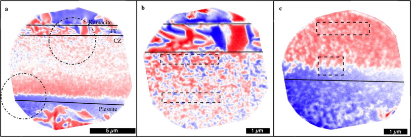
XPEEM Reveals the Detailed Nanomagnetic Structure of the Widmanstätten microstructure in the Tazewell meteorite.
Leave a Reply